Canine histiocytoma is a common benign skin tumor in dogs. It typically presents as a raised, firm, and often ulcerated mass on the head, ears, or limbs. Canine histiocytoma images can be a valuable tool for veterinarians in diagnosing and monitoring this condition.
Histologically, canine histiocytomas are characterized by the presence of plump, round to polygonal cells with abundant eosinophilic cytoplasm and a round to oval nucleus. These cells are often arranged in sheets or nests, and they may contain numerous mitotic figures. The cytoplasm of these cells often contains cytoplasmic vacuoles or inclusions, which can be helpful in distinguishing them from other types of skin tumors.
Canine histiocytomas are typically benign and self-limiting, and they usually regress spontaneously within a few weeks to months. However, in some cases, they may become locally aggressive or even metastasize to other parts of the body. Therefore, it is important for veterinarians to be able to accurately diagnose and monitor canine histiocytomas in order to ensure that they are managed appropriately.
Read also:Leonardo Dicaprios Love Life From Relationships To Marriage
Canine Histiocytoma Images
Canine histiocytoma images are a valuable tool for veterinarians in diagnosing and monitoring this common skin tumor in dogs. These images can help to differentiate canine histiocytomas from other types of skin tumors, and they can also be used to track the progression of the tumor over time.
- Benign
- Self-limiting
- Firm
- Ulcerated
- Head
- Ears
- Limbs
- Histopathology
Canine histiocytomas are typically benign and self-limiting, meaning that they will usually regress spontaneously within a few weeks to months. However, in some cases, they may become locally aggressive or even metastasize to other parts of the body. Therefore, it is important for veterinarians to be able to accurately diagnose and monitor canine histiocytomas in order to ensure that they are managed appropriately.
1. Benign
Benign is a term used to describe a tumor that is not cancerous. Canine histiocytomas are typically benign tumors, meaning that they are not likely to spread to other parts of the body and they are not likely to cause serious health problems.
- Slow-growing
Benign tumors typically grow slowly and do not invade surrounding tissues.
- Well-defined borders
Benign tumors often have well-defined borders, which makes them easy to distinguish from surrounding healthy tissue.
- Do not metastasize
Benign tumors do not metastasize, meaning that they do not spread to other parts of the body.
Read also:
- Insights Into Charles Meltons Wife And Their Relationship
The benign nature of canine histiocytomas is one of the reasons why they are typically not treated. However, if a histiocytoma is causing discomfort or if it is located in an area where it is likely to be irritated, it may be necessary to remove it surgically.
2. Self-limiting
Self-limiting is a term used to describe a condition that will resolve on its own without treatment. Canine histiocytomas are typically self-limiting tumors, meaning that they will usually regress spontaneously within a few weeks to months.
The self-limiting nature of canine histiocytomas is one of the reasons why they are typically not treated. However, if a histiocytoma is causing discomfort or if it is located in an area where it is likely to be irritated, it may be necessary to remove it surgically.
Canine histiocytoma images can be helpful in monitoring the progression of the tumor over time. By comparing images taken at different time points, veterinarians can assess whether the tumor is growing or shrinking. This information can help to determine whether or not treatment is necessary.
3. Firm
Canine histiocytoma images can be useful in assessing the firmness of a tumor. Firmness is a measure of how hard or soft a mass is to the touch. It is an important clinical sign that can help to differentiate between different types of tumors.
- Palpation
Palpation is a physical examination technique used to assess the firmness of a mass. It involves gently feeling the mass with the fingers to determine its consistency.
- Ultrasound
Ultrasound is a medical imaging technique that can be used to assess the firmness of a mass. It involves using sound waves to create an image of the mass.
- Biopsy
A biopsy is a procedure in which a small sample of tissue is removed from the mass and examined under a microscope. This can help to determine the type of tumor and its firmness.
The firmness of a canine histiocytoma can vary depending on the stage of the tumor. Early-stage tumors are typically soft and mobile, while late-stage tumors may be firm and fixed to the underlying tissue.
4. Ulcerated
Ulcerated is a term used to describe a skin lesion that has broken down and formed an open sore. Canine histiocytomas are often ulcerated, which can make them painful and difficult to manage.
There are a number of factors that can contribute to ulceration of canine histiocytomas, including:
- Trauma: Canine histiocytomas are often located on areas of the body that are prone to trauma, such as the head, ears, and limbs. Trauma can cause the tumor to break down and ulcerate.
- Infection: Canine histiocytomas can become infected with bacteria or other microorganisms. This can lead to inflammation and ulceration of the tumor.
- Immune response: The body's immune response to canine histiocytomas can also lead to ulceration. The immune system can attack the tumor cells, causing them to break down and ulcerate.
Ulcerated canine histiocytomas can be a challenge to manage. They can be painful and difficult to keep clean. In some cases, ulcerated histiocytomas may need to be treated with antibiotics or other medications to prevent infection. Surgery may also be necessary to remove the tumor if it is causing significant problems.
Canine histiocytoma images can be helpful in assessing the severity of ulceration. By comparing images taken at different time points, veterinarians can determine whether the ulceration is getting worse or better. This information can help to guide treatment decisions.
5. Head
The head is one of the most common locations for canine histiocytomas. These tumors often appear on the face, ears, or neck. There are a number of reasons why the head is a common site for histiocytomas:
- Abundant sun exposure
The head is often exposed to the sun, which can damage the skin and lead to the development of histiocytomas.
- Trauma
The head is also a common site for trauma, which can further increase the risk of developing histiocytomas.
- Immune response
The immune system is responsible for fighting off infection and disease. However, the immune system can also attack healthy cells, which can lead to the development of histiocytomas.
Canine histiocytoma images can be helpful in diagnosing and monitoring histiocytomas on the head. By comparing images taken at different time points, veterinarians can assess whether the tumor is growing or shrinking. This information can help to determine whether or not treatment is necessary.
6. Ears
The ears are another common location for canine histiocytomas. These tumors often appear on the ear pinna, which is the visible part of the ear. In some cases, histiocytomas may also develop in the ear canal.
There are a number of reasons why the ears are a common site for histiocytomas:
- Abundant sun exposure
The ears are often exposed to the sun, which can damage the skin and lead to the development of histiocytomas.
- Trauma
The ears are also a common site for trauma, which can further increase the risk of developing histiocytomas.
- Immune response
The immune system is responsible for fighting off infection and disease. However, the immune system can also attack healthy cells, which can lead to the development of histiocytomas.
Canine histiocytoma images can be helpful in diagnosing and monitoring histiocytomas on the ears. By comparing images taken at different time points, veterinarians can assess whether the tumor is growing or shrinking. This information can help to determine whether or not treatment is necessary.
7. Limbs
The limbs are a common location for canine histiocytomas. These tumors can occur on any part of the limb, including the upper leg, lower leg, paw, and toes. There are a number of reasons why the limbs are a common site for histiocytomas:
- Trauma
The limbs are often exposed to trauma, which can damage the skin and lead to the development of histiocytomas.
- Sun exposure
The limbs are also often exposed to the sun, which can further increase the risk of developing histiocytomas.
- Immune response
The immune system is responsible for fighting off infection and disease. However, the immune system can also attack healthy cells, which can lead to the development of histiocytomas.
Canine histiocytoma images can be helpful in diagnosing and monitoring histiocytomas on the limbs. By comparing images taken at different time points, veterinarians can assess whether the tumor is growing or shrinking. This information can help to determine whether or not treatment is necessary.
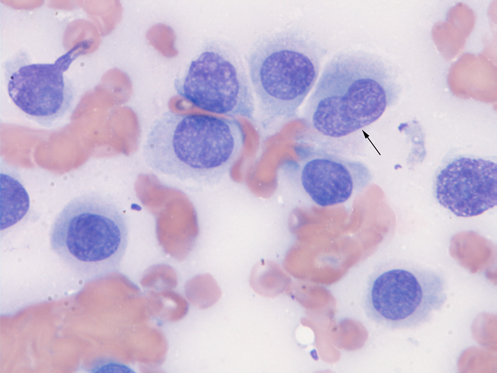
8. Histopathology
Histopathology is the study of tissues under a microscope. It is an essential tool for diagnosing and classifying diseases, including canine histiocytomas. Canine histiocytoma images can provide valuable information for histopathologists, as they can help to identify the characteristic features of these tumors.
- Cellular composition
Canine histiocytomas are composed of a variety of cells, including histiocytes, lymphocytes, and neutrophils. The relative proportions of these cells can vary depending on the stage of the tumor.
- Architectural patterns
Canine histiocytomas can exhibit a variety of architectural patterns, including sheets, nests, and cords. The arrangement of the cells can provide clues to the aggressiveness of the tumor.
- Mitotic activity
Mitotic activity is a measure of how quickly a tumor is growing. Canine histiocytomas typically have a low mitotic rate, which indicates that they are slow-growing tumors.
- Necrosis
Necrosis is the death of tissue. It can be a sign of a rapidly growing tumor. Canine histiocytomas typically do not exhibit necrosis.
Canine histiocytoma images can be used to assess all of these histopathologic features. This information can help pathologists to make a definitive diagnosis of canine histiocytoma and to determine the best course of treatment.
FAQs on Canine Histiocytoma Images
Canine histiocytoma images are a valuable tool for veterinarians in diagnosing and monitoring this common skin tumor in dogs. Here are some frequently asked questions about canine histiocytoma images:
Question 1: What are the benefits of using canine histiocytoma images?
Answer: Canine histiocytoma images can help veterinarians to:
- Diagnose canine histiocytomas
- Monitor the progression of canine histiocytomas
- Differentiate canine histiocytomas from other types of skin tumors
- Determine the best course of treatment for canine histiocytomas
Question 2: How are canine histiocytoma images obtained?
Answer: Canine histiocytoma images are typically obtained using a technique called cytology. Cytology involves collecting a sample of cells from the tumor and examining them under a microscope.
Question 3: What do canine histiocytoma images look like?
Answer: Canine histiocytoma images typically show a collection of round or polygonal cells with abundant cytoplasm and a round or oval nucleus. The cells may be arranged in sheets, nests, or cords.
Question 4: How can canine histiocytoma images help to diagnose canine histiocytomas?
Answer: Canine histiocytoma images can help to diagnose canine histiocytomas by showing the characteristic features of these tumors, such as the presence of round or polygonal cells with abundant cytoplasm and a round or oval nucleus. These features can help to differentiate canine histiocytomas from other types of skin tumors.
Question 5: How can canine histiocytoma images help to monitor the progression of canine histiocytomas?
Answer: Canine histiocytoma images can help to monitor the progression of canine histiocytomas by showing changes in the size, shape, and appearance of the tumor over time. This information can help veterinarians to determine whether the tumor is growing or shrinking and whether treatment is necessary.
Question 6: How can canine histiocytoma images help to determine the best course of treatment for canine histiocytomas?
Answer: Canine histiocytoma images can help to determine the best course of treatment for canine histiocytomas by showing the characteristics of the tumor. This information can help veterinarians to determine whether the tumor is likely to be benign or malignant and whether surgery, radiation therapy, or chemotherapy is the best course of treatment.
Canine histiocytoma images are a valuable tool for veterinarians in diagnosing and managing canine histiocytomas. By providing a visual representation of the tumor, canine histiocytoma images can help veterinarians to make more accurate diagnoses, monitor the progression of the tumor, and determine the best course of treatment.
Tips on Using Canine Histiocytoma Images
Canine histiocytoma images are a valuable tool for veterinarians in diagnosing and managing canine histiocytomas. Here are some tips on how to use canine histiocytoma images effectively:
Tip 1: Use high-quality images.
The quality of the images you use will have a significant impact on your ability to diagnose and monitor canine histiocytomas. Make sure to use high-resolution images that are in focus and well-lit.
Tip 2: Take multiple images.
Taking multiple images of the tumor from different angles can help you to get a better understanding of its size, shape, and location. This information can be helpful in diagnosing the tumor and determining the best course of treatment.
Tip 3: Compare images over time.
Comparing images of the tumor taken over time can help you to monitor its progression. This information can help you to determine whether the tumor is growing or shrinking and whether treatment is necessary.
Tip 4: Share images with your veterinarian.
Sharing images of the tumor with your veterinarian can help them to make a more accurate diagnosis and determine the best course of treatment. Your veterinarian may also be able to provide you with additional information about canine histiocytomas and their treatment.
Tip 5: Use images to educate yourself.
Canine histiocytoma images can be a valuable tool for educating yourself about canine histiocytomas. By studying images of these tumors, you can learn more about their appearance, behavior, and treatment.
Canine histiocytoma images are a powerful tool that can help you to diagnose, monitor, and treat canine histiocytomas. By following these tips, you can use canine histiocytoma images effectively to improve the health of your dog.