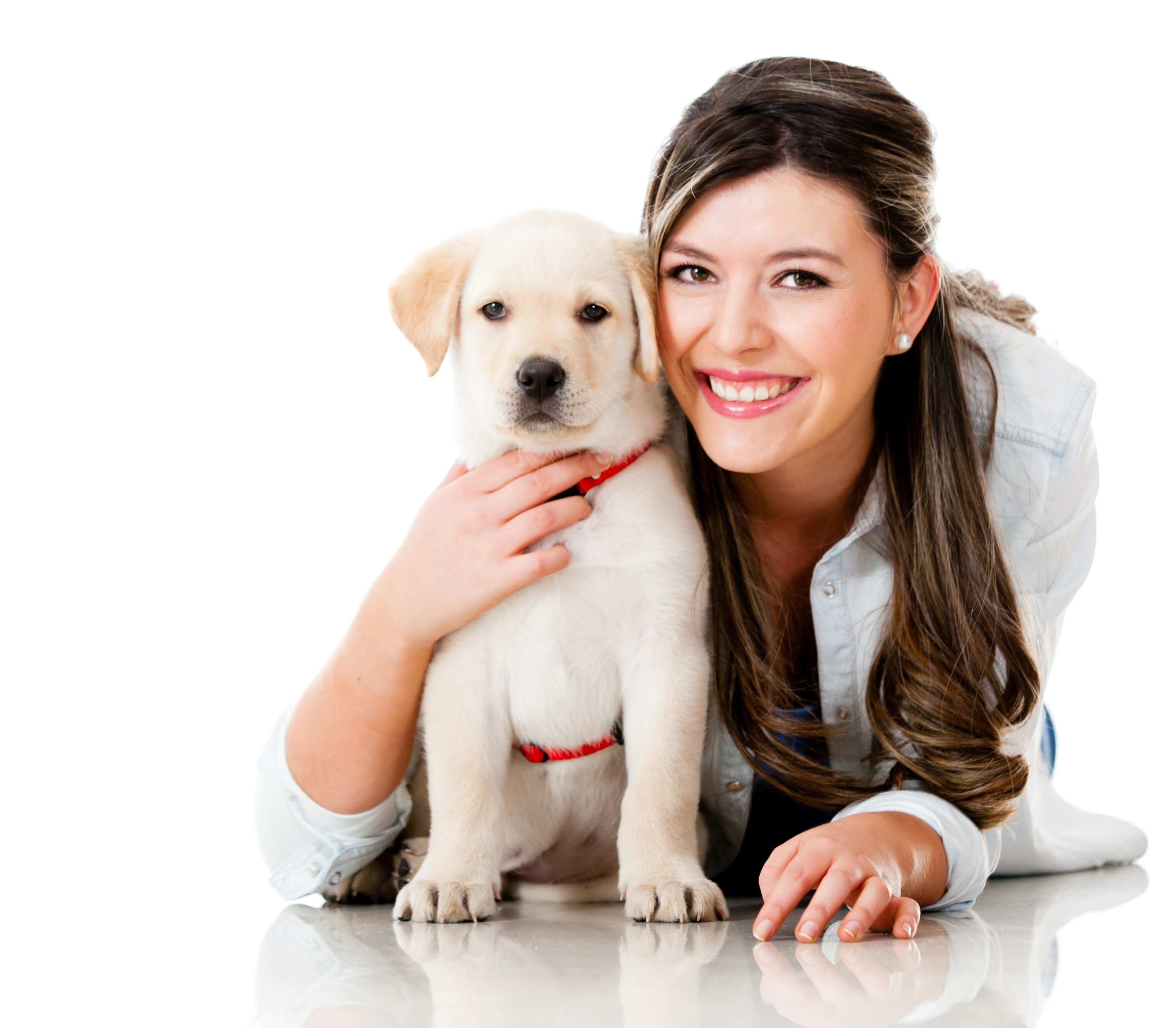
Histiocytoma is a benign skin tumor that is most commonly seen in young dogs. It is usually a small, raised, red or pink bump that can appear anywhere on the body. Histiocytomas are typically not painful or itchy, and they usually disappear on their own within a few months. However, if the histiocytoma is large or bothersome, it can be removed surgically.
Images of histiocytoma in dogs can be helpful for dog owners to identify and track the progress of this common skin tumor. These images can also be used by veterinarians to diagnose histiocytoma and to rule out other more serious conditions.
In this article, we will provide a comprehensive overview of histiocytoma in dogs, including information on its symptoms, diagnosis, treatment, and prognosis. We will also provide a number of images of histiocytoma in dogs to help you identify this condition.
Read also:Unraveling Vinny Guadagninos Relationship Status And Personal Life
Images of Histiocytoma in Dogs
Images of histiocytoma in dogs are an important tool for dog owners and veterinarians to identify and track this common skin tumor. These images can help to differentiate histiocytoma from other more serious conditions, and they can also be used to monitor the progress of treatment.
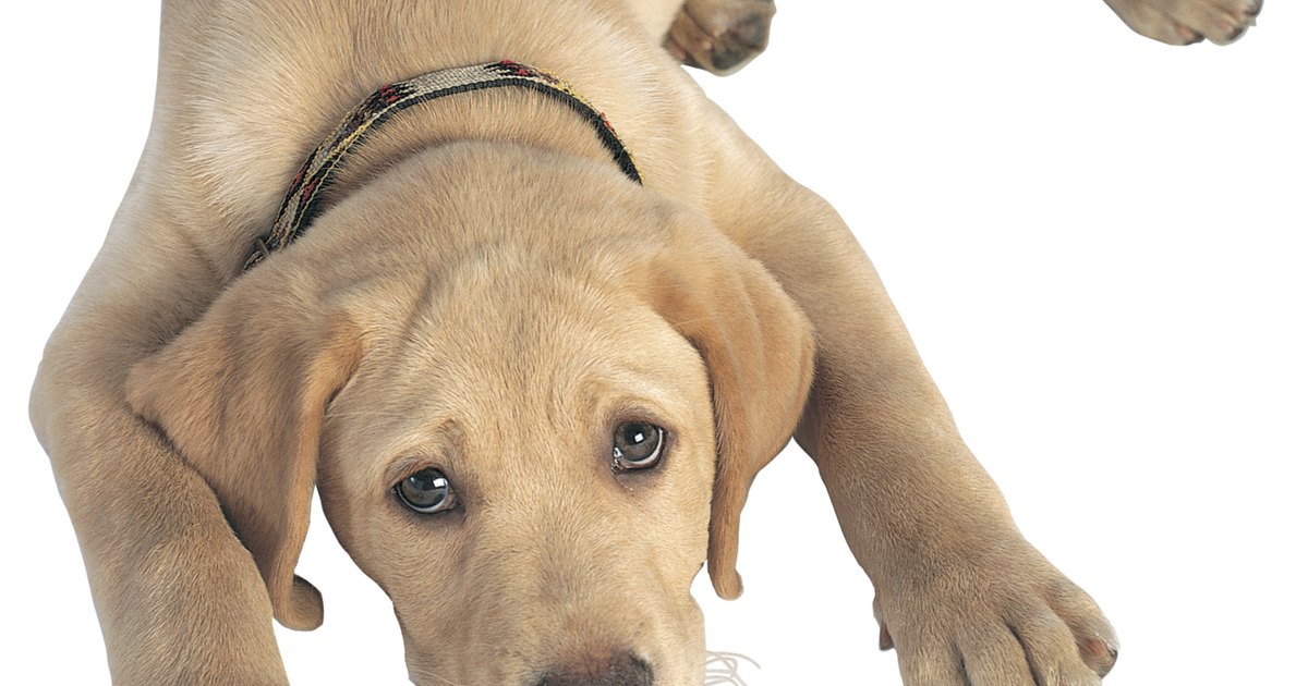
- Clinical Appearance: Histiocytomas are typically small, raised, red or pink bumps that can appear anywhere on the body.
- Age of Onset: Histiocytomas are most commonly seen in young dogs, between the ages of 1 and 3.
- Breed Predisposition: Histiocytomas are more common in certain breeds of dogs, such as Boxers, Bulldogs, and Rottweilers.
- Diagnosis: Histiocytomas are typically diagnosed based on their clinical appearance and history. However, a biopsy may be necessary to rule out other conditions.
- Treatment: Histiocytomas usually disappear on their own within a few months. However, if the histiocytoma is large or bothersome, it can be removed surgically.
- Prognosis: Histiocytomas are typically benign and do not spread to other parts of the body. However, in rare cases, histiocytomas can become malignant.
Images of histiocytoma in dogs can be a valuable resource for dog owners and veterinarians. These images can help to identify and track this common skin tumor, and they can also be used to rule out other more serious conditions.
1. Clinical Appearance
The clinical appearance of histiocytoma is an important factor in diagnosing this common skin tumor in dogs. Histiocytomas are typically small, raised, red or pink bumps that can appear anywhere on the body. This characteristic appearance is often enough for a veterinarian to make a diagnosis of histiocytoma, but in some cases, a biopsy may be necessary to rule out other conditions.
Images of histiocytoma in dogs can be a valuable tool for dog owners and veterinarians to identify and track this condition. These images can help to differentiate histiocytoma from other more serious conditions, such as mast cell tumors or melanomas. Additionally, images of histiocytoma can be used to monitor the progress of treatment and to ensure that the tumor is not spreading.
In summary, the clinical appearance of histiocytoma is an important factor in diagnosing and managing this condition. Images of histiocytoma in dogs can be a valuable tool for dog owners and veterinarians to identify, track, and treat this common skin tumor.
2. Age of Onset
There is a strong connection between the age of onset of histiocytoma and the appearance of this condition in images of histiocytoma in dogs. Histiocytomas are most commonly seen in young dogs, between the ages of 1 and 3. This is because the immune system of young dogs is still developing, and they are more likely to develop histiocytomas as a reaction to insect bites, vaccinations, or other irritants.
Read also:Clytie Lane The Intriguing Life And Influence Of A Remarkable Personality
Images of histiocytoma in dogs can help to confirm the diagnosis of this condition. In young dogs, the presence of a small, raised, red or pink bump is highly suggestive of histiocytoma. However, in older dogs, histiocytomas may be more difficult to diagnose, as they may appear differently and may be more likely to be mistaken for other conditions, such as mast cell tumors or melanomas.
Therefore, it is important to be aware of the age of onset of histiocytoma when interpreting images of histiocytoma in dogs. This information can help to narrow down the diagnosis and to ensure that the most appropriate treatment is provided.
3. Breed Predisposition
The breed of a dog is an important factor to consider when interpreting images of histiocytoma in dogs. Certain breeds of dogs are more predisposed to developing histiocytomas than others. These breeds include Boxers, Bulldogs, and Rottweilers.
- Genetic Factors: Histiocytomas are thought to be caused by a combination of genetic and environmental factors. Certain breeds of dogs may have a genetic predisposition to developing histiocytomas. This is because they may have a weakened immune system or a higher sensitivity to certain environmental triggers.
- Environmental Triggers: Histiocytomas are often triggered by environmental factors, such as insect bites, vaccinations, or other irritants. These triggers can cause the immune system to overreact and produce histiocytomas.
- Clinical Appearance: Histiocytomas in certain breeds of dogs may have a characteristic clinical appearance. For example, histiocytomas in Boxers are often multiple and may appear on the face and legs. Histiocytomas in Bulldogs are often solitary and may appear on the head and neck.
- Treatment and Prognosis: The treatment and prognosis of histiocytoma in dogs may vary depending on the breed of the dog. For example, histiocytomas in Boxers are often more aggressive and may require more extensive treatment than histiocytomas in other breeds.
Therefore, it is important to be aware of the breed predisposition of histiocytoma when interpreting images of histiocytoma in dogs. This information can help to narrow down the diagnosis and to ensure that the most appropriate treatment is provided.
4. Diagnosis
Images of histiocytoma in dogs can play a crucial role in the diagnosis of this common skin tumor. By providing a visual representation of the tumor's characteristics, images can help veterinarians to make a more accurate diagnosis and to rule out other conditions that may have a similar appearance.
- Clinical Appearance: The clinical appearance of histiocytoma is often enough for a veterinarian to make a diagnosis. Histiocytomas are typically small, raised, red or pink bumps that can appear anywhere on the body. However, in some cases, the clinical appearance of histiocytoma may be similar to other conditions, such as mast cell tumors or melanomas.
- History: The history of the tumor can also be helpful in making a diagnosis of histiocytoma. Histiocytomas are most commonly seen in young dogs, between the ages of 1 and 3. Additionally, certain breeds of dogs are more predisposed to developing histiocytomas than others.
- Biopsy: In some cases, a biopsy may be necessary to rule out other conditions that may have a similar appearance to histiocytoma. A biopsy involves removing a small sample of the tumor and examining it under a microscope.
Images of histiocytoma in dogs can be a valuable tool for veterinarians to diagnose this common skin tumor. By providing a visual representation of the tumor's characteristics, images can help to make a more accurate diagnosis and to rule out other conditions that may have a similar appearance.
5. Treatment
Images of histiocytoma in dogs can provide valuable information about the size and location of the tumor, which can be helpful in determining the best course of treatment. In most cases, histiocytomas will disappear on their own within a few months. However, if the histiocytoma is large or bothersome, it can be removed surgically.
There are several factors to consider when making the decision of whether or not to remove a histiocytoma surgically. These factors include the size and location of the tumor, the age and health of the dog, and the owner's financial situation.
If the histiocytoma is small and located in a non-bothersome area, it may be best to simply monitor the tumor and wait for it to disappear on its own. However, if the histiocytoma is large or located in a bothersome area, such as on the face or near the eyes, it may be necessary to remove it surgically.
The surgical removal of a histiocytoma is a relatively simple and straightforward procedure. The surgery is typically performed under local anesthesia, and the tumor is removed with a scalpel. The incision is then closed with stitches, and the dog is sent home the same day.
The prognosis for dogs with histiocytoma is generally good. Most histiocytomas will disappear on their own within a few months, and even those that require surgical removal are unlikely to recur.
6. Prognosis
Images of histiocytoma in dogs can provide valuable information about the prognosis of this condition. In most cases, histiocytomas are benign and do not spread to other parts of the body. However, in rare cases, histiocytomas can become malignant.
The prognosis for dogs with malignant histiocytoma is generally poor. These tumors are often aggressive and difficult to treat. However, early diagnosis and treatment can improve the prognosis.
If you are concerned that your dog may have a histiocytoma, it is important to take them to the veterinarian for a diagnosis. The veterinarian will be able to examine the tumor and determine if it is benign or malignant. If the tumor is malignant, the veterinarian will recommend the best course of treatment.
Here are some key insights to remember about the prognosis of histiocytoma in dogs:
- Most histiocytomas are benign and do not spread to other parts of the body.
- In rare cases, histiocytomas can become malignant.
- The prognosis for dogs with malignant histiocytoma is generally poor.
- Early diagnosis and treatment can improve the prognosis.
FAQs About Images of Histiocytoma in Dogs
Images of histiocytoma in dogs can be a valuable resource for dog owners and veterinarians to identify and track this common skin tumor. These images can help to differentiate histiocytoma from other more serious conditions, and they can also be used to monitor the progress of treatment.
Question 1: What is a histiocytoma?
Answer 1: A histiocytoma is a benign skin tumor that is most commonly seen in young dogs. It is usually a small, raised, red or pink bump that can appear anywhere on the body.
Question 2: How are histiocytomas diagnosed?
Answer 2: Histiocytomas are typically diagnosed based on their clinical appearance and history. However, a biopsy may be necessary to rule out other conditions.
Question 3: What is the treatment for histiocytomas?
Answer 3: Histiocytomas usually disappear on their own within a few months. However, if the histiocytoma is large or bothersome, it can be removed surgically.
Question 4: What is the prognosis for dogs with histiocytoma?
Answer 4: The prognosis for dogs with histiocytoma is generally good. Most histiocytomas will disappear on their own within a few months, and even those that require surgical removal are unlikely to recur.
Question 5: Are there any breeds of dogs that are more likely to develop histiocytomas?
Answer 5: Yes, certain breeds of dogs, such as Boxers, Bulldogs, and Rottweilers, are more likely to develop histiocytomas.
Question 6: Can histiocytomas become malignant?
Answer 6: In rare cases, histiocytomas can become malignant. The prognosis for dogs with malignant histiocytoma is generally poor.
Summary of key takeaways or final thought: Images of histiocytoma in dogs can be a valuable resource for dog owners and veterinarians to identify and track this common skin tumor. These images can help to differentiate histiocytoma from other more serious conditions, and they can also be used to monitor the progress of treatment.
Transition to the next article section: If you are concerned that your dog may have a histiocytoma, it is important to take them to the veterinarian for a diagnosis. The veterinarian will be able to examine the tumor and determine if it is benign or malignant. If the tumor is malignant, the veterinarian will recommend the best course of treatment.
Tips for Identifying and Treating Histiocytoma in Dogs Using Images
Images of histiocytoma in dogs can be a valuable tool for dog owners and veterinarians to identify and track this common skin tumor. These images can help to differentiate histiocytoma from other more serious conditions, and they can also be used to monitor the progress of treatment.
Tip 1:Take clear and well-lit photos of the histiocytoma.
This will help the veterinarian to get a good look at the tumor and to make a more accurate diagnosis.
Tip 2:Take photos of the histiocytoma from different angles.
This will give the veterinarian a better understanding of the tumor's size and location.
Tip 3:Note the size and location of the histiocytoma.
This information can be helpful in determining the best course of treatment.
Tip 4:Monitor the histiocytoma for changes.
If the tumor changes in size, shape, or color, it is important to take the dog to the veterinarian for a re-examination.
Tip 5:Follow the veterinarian's instructions for treatment.
This may include topical medications, surgery, or other treatments.
Summary of key takeaways or benefits: By following these tips, dog owners and veterinarians can use images of histiocytoma in dogs to identify and track this common skin tumor. This information can help to ensure that the dog receives the best possible care.
Transition to the article's conclusion: If you are concerned that your dog may have a histiocytoma, it is important to take them to the veterinarian for a diagnosis. The veterinarian will be able to examine the tumor and determine if it is benign or malignant. If the tumor is malignant, the veterinarian will recommend the best course of treatment.
Conclusion
Images of histiocytoma in dogs are a valuable tool for dog owners and veterinarians to identify and track this common skin tumor. These images can help to differentiate histiocytoma from other more serious conditions, and they can also be used to monitor the progress of treatment.
By understanding the clinical appearance, age of onset, breed predisposition, diagnosis, treatment, and prognosis of histiocytoma, dog owners and veterinarians can work together to ensure that dogs with this condition receive the best possible care.